


E 5-15% pea o ngā whati koi katoa o te wheua navicular ka puta te navicular malunion, ā, e 3% pea te hunga ka pāngia e te navicular necrosis. Ko ngā āhuatanga mōrearea mō te navicular malunion ko te korenga o te tātaritanga, te tata o te rārangi whati, te nekenga neke atu i te 1 mm, me te whati me te kore pumau o te carpal. Mēnā kāore e rongoatia, he maha ngā wā ka honoa te navicular osteochondral nonunion ki te traumatic arthritis, e mōhiotia ana ko te navicular osteochondral nonunion me te collapsing osteoarthritis.
Ka taea te whakamahi i te honohono wheua me te kore rānei he papa kua whakapūmauhia hei rongoā i te navicular osteochondral nonunion. Heoi, mō ngā tūroro e pāngia ana e te osteonecrosis o te pou proximal o te wheua navicular, kāore i te pai ngā hua o te honohono wheua me te kore he pito vascular, ā, ko te tere whakaora wheua he 40%-67% noa iho. He rerekē, ko te tere whakaora o ngā honohono wheua me ngā papa kua whakapūmauhia ka taea te teitei ki te 88%-91%. Ko ngā papa wheua kua whakapūmauhia nui i roto i te mahi haumanu ko te 1,2-ICSRA-tipped distal radius flap, te bone graft + vascular bundle implant, te palmar radius flap, te free iliac bone flap me te pito kua whakapūmauhia, me te medial femoral condylar bone flap (MFC VBG), me ētahi atu. He pai ngā hua o te honohono wheua me te pito kua whakapūmauhia. Kua whakaatuhia te whai huatanga o te free MFC VBG ki te rongoā i ngā whati navicular me te metacarpal collapse, ā, ka whakamahia e te MFC VBG te peka hononga o te uaua turi heke hei peka trophic matua. Ki te whakaritea ki ētahi atu papa, ka whakaratohia e te MFC VBG he tautoko hanganga e ranea ana hei whakahoki mai i te āhua noa o te wheua navicular, inā koa i roto i te osteochondrosis whati navicular me te piko o te tuara (Pikitia 1). I roto i te maimoatanga o te osteonecrosis osteochondral navicular me te hinganga o te carpal, kua pūrongohia he 40% noa iho te tere whakaora wheua o te papa radius distal 1,2-ICSRA-tipped, engari he 100% te tere whakaora wheua o te MFC VBG.
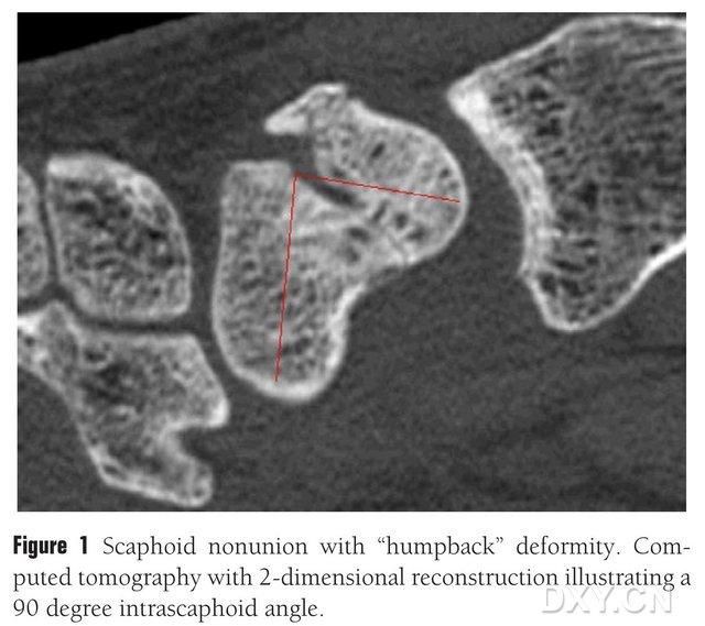
Pikitia 1. Te whati o te wheua navicular me te āhua "piko ki muri", e whakaatu ana te CT i te poraka whati i waenganui i ngā wheua navicular i te koki o te 90° pea.
Te whakarite i mua i te pokanga
I muri i te whakamātautau tinana o te ringaringa e pāngia ana, me whakahaere he rangahau whakaahua hei aromatawai i te nui o te hinga o te ringaringa. He pai ngā whakaahua X-ray māmā hei whakaū i te wāhi o te whati, te nui o te nekehanga, me te aroaro o te mimiti, te sclerosis rānei o te pito whati. Ka whakamahia ngā whakaahua o muri o mua hei aromatawai i te hinga o te ringaringa, te kore pumau o te hope o te ringaringa (DISI) mā te whakamahi i te ōwehenga teitei o te ringaringa kua whakarerekētia (teitei/whānui) o ≤1.52, i te koki radial lunate neke atu rānei i te 15°. Ka taea e te MRI, te CT rānei te āwhina ki te tautuhi i te kore tika o te wheua navicular, te osteonecrosis rānei. Ko ngā whakaahua X-ray taha, ko te CT sagittal oblique rānei o te wheua navicular me te koki navicular >45° e tohu ana i te whakapoto o te wheua navicular, e mōhiotia ana ko te "bowed back deformity". Ko te tohu MRI T1, T2 iti e tohu ana i te nekrosis o te wheua navicular, engari kāore he hiranga mārama o te MRI ki te whakatau i te whakaora o te whati.
Ngā tohu me ngā contraindications:
Ko te kore-huinga o te osteochondral navicular me te piko o te tuara me te DISI; e whakaatu ana te MRI i te necrosis ischemic o te wheua navicular, te wetewete o te tourniquet i te wā o te pokanga me te tirotiro i te pito whati o te wheua navicular he wheua sclerotic ma tonu; ko te korenga o te hononga wheua wedge tuatahi, te whakapiringa ā-roto rānei me whai hononga wheua hanganga VGB nui (>1cm3). ngā kitenga i mua i te pokanga, i te wā rānei o te pokanga o te osteoarthritis o te hononga carpal radial; mena kua puta he malunion navicular nui me te osteoarthritis hinga, ka hiahiatia pea te tango i te nerve o te ringaringa, te osteotomy navicular, te whakakotahitanga quadrangular, te osteotomy carpal proximal, te whakakotahitanga carpal katoa, me ētahi atu; malunion navicular, necrosis proximal, engari me te āhua noa o te wheua navicular (hei tauira, te whati navicular kore-neke me te kore toto e rere ki te pou proximal); te whakapoto o te malunion navicular me te kore osteonecrosis. (Ka taea te whakamahi i te 1,2-ICSRA hei whakakapi mō te flap radius distal).
Anatomy Whakamahia
Ko te MFC VBG e tukuna ana e te maha o ngā oko toto trophoblastic iti i waenganui i ngā wheua (te toharite 30, 20-50), ko te nuinga o te toto e tukuna ana ki te taha muri o raro i te condyle femoral medial (te toharite 6.4), ā, muri iho ko te taha mua o runga (te toharite 4.9) (Pikitia 2). Ko te nuinga o te toto e tukuna ana ki ēnei oko toto trophoblastic ko te uaua geniculate descending (DGA) me/ranei te uaua geniculate medial superior (SMGA), he peka o te uaua femoral superficial e puta ai hoki ngā peka io articular, musculocutaneous, me/ranei ngā peka io saphenous. I ahu mai te DGA i te uaua femoral superficial proximal ki te medial eminence o te malleolus medial, i te tawhiti rānei o te 13.7 cm proximal ki te mata articular (10.5-17.5 cm), ā, ko te pumau o te peka he 89% i roto i ngā tauira tupapaku (Pikitia 3). Ka ahu mai te DGA i te uaua femoral o runga i te 13.7 cm (10.5 cm-17.5 cm) e tata ana ki te kapiti malleolus medial, e tata ana rānei ki te mata hononga, me te tauira tūpāpaku e whakaatu ana i te pumau o te manga 100% me te diameter o te 0.78 mm pea. Nō reira, e whakaaetia ana te DGA, te SMGA rānei, ahakoa he pai ake te mea tuatahi mō ngā tibiae nā te roa me te diameter o te uaua.
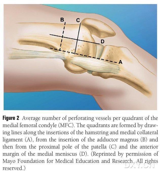
Pikitia 2. Te tohatoha o ngā oko toto trophoblast MFC i te rārangi whakapae i waenganui i te semitendinosus me te ligament collateral medial A, te rārangi o te trochanter nui B, te rārangi o te pou o runga o te patella C, te rārangi o te meniscus o mua D.
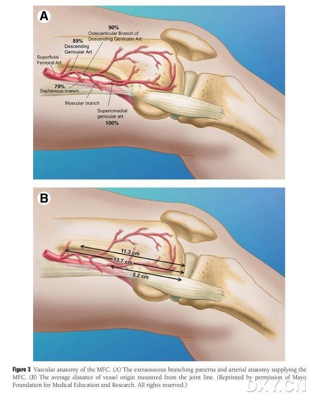
Pikitia 3. Te hanganga o ngā oko toto MFC: (A) Ngā manga extraosseous me te hanganga o ngā oko toto trophoblastic MFC, (B) Te tawhiti o ngā pūtake oko toto mai i te rārangi hononga
Te urunga pokanga
Ka whakanohoia te tūroro ki raro i te rongoā whakamoe whānui i te tūnga takoto, me te waewae e pāngia ana e whakatakotoria ana ki runga i te tēpu pokanga ringa. Ko te tikanga, ka tangohia te kiri wheua kaituku mai i te condyle femoral medial ipsilateral, kia taea ai e te tūroro te neke me ngā tokotoko i muri i te pokanga. Ka taea hoki te whiriwhiri i te turi contralateral mēnā he hītori whara, he pokanga rānei i te taha kotahi o te turi. Ka piko te turi, ā, ka hurihia te hope ki waho, ā, ka whakamahia ngā tourniquets ki ngā pito o runga me raro. Ko te huarahi pokanga ko te huarahi Russe whānui, me te tapahi e tīmata ana i te 8 cm te tata ki te kauhanga carpal whakawhiti me te toro atu ki tawhiti mai i te taha radial o te uaua radial flexor carpi radialis, kātahi ka piko i te kauhanga carpal whakawhiti ki te turanga o te koromatua, ka mutu ki te taumata o te trochanter nui. Ka tapahia te anga o te uaua o te uaua radial longissimus, ā, ka tōia te uaua ki te taha maui, ā, ka kitea te wheua navicular mā te tapahi koi i te taha o ngā hononga radial lunate me te radial navicular head, me te wehe āta i ngā kiko ngohengohe o te wheua navicular kia kitea ai te wheua navicular (Pikitia 4). Whakaūtia te rohe o te kore-hononga, te kounga o te kiriuhi hononga me te nui o te ischemia o te wheua navicular. Whai muri i te wetewete i te tourniquet, tirohia te pou proximal o te wheua navicular mō te toto punctate hei whakatau mēnā he ischaemic necrosis. Mena kāore te navicular necrosis e pā ana ki te radial carpal, intercarpal arthritis rānei, ka taea te whakamahi i te MFC VGB.
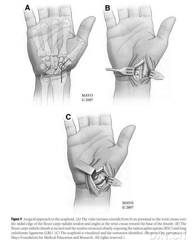
Pikitia 4. Te huarahi pokanga navicular: (A) Ka tīmata te tapahi i te 8 cm te tawhiti atu ki te kauhanga carpal whakawhiti, ā, ka toro atu i te taha radial o te uaua radial flexor carpi radialis ki te wāhanga tawhiti o te tapahi, e piko ana ki te turanga o te koromatua i te kauhanga carpal whakawhiti. (B) Ka tapahia te anga uaua o te uaua radial longissimus, ā, ka kumea te uaua ki te taha ulnar, ā, ka whakaatuhia te wheua navicular mā te tapahi koi i te taha o ngā hononga radial lunate me te radial navicular head. (C) Tāutuhia te rohe o te motuhanga wheua navicular.
Ka tapahia he tapahi 15-20 cm te roa ki te taha o muri o te uaua femur o waenganui, ā, ka hoki whakamuri te uaua ki mua kia kitea ai te toto MFC (Pikitia 5). Ko ngā peka hononga o te DGA me te SMGA te nuinga o te wā e tukuna ana te toto MFC, ko te nuinga o te wā ka tangohia te peka hononga nui o te DGA me te uaua e haere tahi ana. Ka tukuna te pedicle vascular ki te taha o muri, me te tiaki i te periosteum me ngā oko toto trophoblastic i runga i te mata wheua.
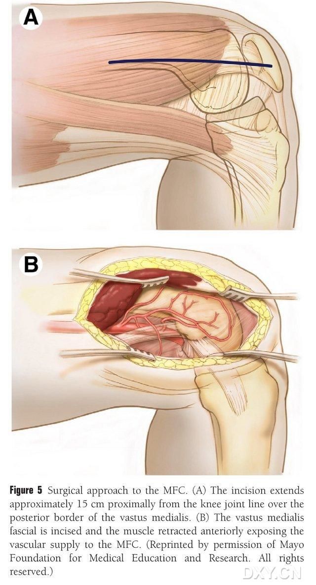
Pikitia 5. Te urunga pokanga ki te MFC: (A) Ka tapahia he tapahi 15-20 cm te roa ki te taha o muri o te uaua femur medial mai i te hononga turi. (B) Ka hoki whakamuri te uaua ki mua kia kitea ai te rere o te toto MFC.
Te whakarite i te wheua navicular
Me whakatika te āhua o te navicular DISI, ā, me whakarite te horahanga o te hononga wheua osteochondral i mua i te whakaurunga mā te piko o te ringaringa i raro i te fluoroscopy hei whakahoki mai i te koki radial lunate noa (Pikitia 6). Ka pokaia he pine Kirschner 0.0625-putu (tata ki te 1.5-mm) mā te kiri mai i te tuara ki te metacarpal hei whakau i te hononga radial lunate, ā, ka kitea te āputa navicular malunion ina whakatikatikaina te ringaringa. I whakakorea te kiko ngohengohe i te wāhi whati, ā, i tautokona anō ki te horapa pereti. Ka whakamahia he kani iti hei whakawhāiti i te wheua, kia rite ai te papa o te whakaurunga ki te hanganga tapawhā kaua ki te poro, e hiahia ana kia whānui ake te āputa navicular i te taha nikau i te taha tuara. Whai muri i te whakatuwheratanga o te āputa, ka inehia te hapa i roto i ngā āhuatanga e toru hei whakatau i te whānuitanga o te hononga wheua, he 10-12 mm te roa i ngā taha katoa o te hononga.
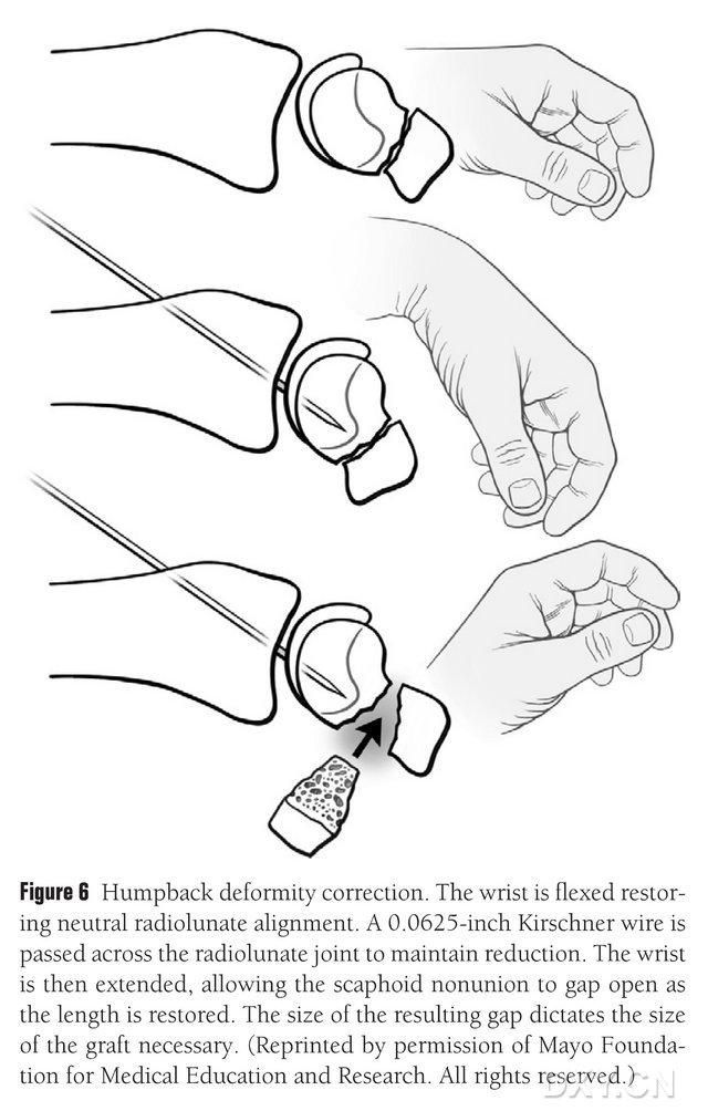
Pikitia 6. Te whakatikatika i te piko o te tuara o te navicular, me te piko o te ringaringa ki te whakamahi i te fluoroscopic hei whakahoki mai i te tūnga radial-lunar noa. Ka pokaia he pine Kirschner 0.0625-putu (tata ki te 1.5-mm) mā te kiri mai i te tuara ki te metacarpal hei whakau i te hononga radial lunate, e whakaatu ana i te āputa navicular malunion me te whakahoki mai i te teitei noa o te wheua navicular ina whakatikatikahia te ringaringa, me te rahi o te āputa e tohu ana i te rahi o te papa e hiahia ana kia āraia.
Te tango wheua
Ko te wāhi kua whakapūpūhia ngā uaua o te condyle femoral medial ka tīpakohia hei wāhi tango wheua, ā, ka tohua pai te wāhi tango wheua. Kia tūpato kei whara te hononga collateral medial. Ka tapahia te periosteum, ā, ka tapahia he parirau wheua tapawhā rite te rahi mō te parirau e hiahiatia ana ki te kani reciprocating, me te tapahi i tētahi poraka wheua tuarua i te 45° i te taha kotahi hei whakarite i te pumau o te parirau (Pikitia 7). 7). Kia tūpato kei wehea te periosteum, te wheua cortical, me te wheua cancellous o te parirau. Me tuku te tourniquet o te pito o raro hei tirotiro i te rere o te toto i roto i te parirau, ā, me tuku te pedicle vascular ki te taha tata mō te iti rawa 6 cm kia taea ai te anastomosis vascular i muri mai. Mena e tika ana, ka taea te haere tonu i tētahi iti o te wheua cancellous i roto i te condyle femoral. Ka whakakīia te hapa condylar femoral ki tētahi whakakapinga wheua, ā, ka tātarihia te tapahi ka kati papatahi i te papatahi.
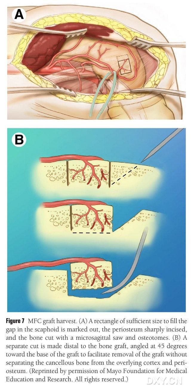
Pikitia 7. Te tango i te papa wheua MFC. (A) Ka tohua te wāhi osteotomy e ranea ana hei whakakī i te wāhi navicular, ka tapahia te periosteum, ā, ka tapahia he papa wheua tapawhā rite te rahi mō te papa e hiahiatia ana ki te kani reciprocating. (B) Ka tapahia he wahi wheua tuarua i te taha kotahi i te 45° hei whakarite i te pumau o te papa.
Te whakaurunga me te whakapiri i te papa
Ka tapahia te papa wheua ki te āhua e tika ana, me te āta tupato kia kaua e pēhia te pedicle vascular, kia kore ai hoki e tangohia te periosteum. Ka āta whakatōkia te papa ki te wāhi o te koha o te wheua navicular, me te karo i te patupatu, ā, ka mau ki ngā tīwiri navicular tuwhera. I āta whakarite kia ōrite te taha nikau o te poraka wheua kua whakatōkia ki te taha nikau o te wheua navicular, kia paku pēhia rānei kia kore ai e pā. I mahia te fluoroscopy hei whakaū i te āhua o te wheua navicular, te rārangi kaha me te tūnga tīwiri. Whakapirihia te uaua papa vascular ki te pito o te uaua radial ki te taha, me te pito venous ki te uaua hoa o te uaua radial mai i te pito ki te pito (Pikitia 8). Ka whakatikahia te capsule hononga, engari ka karohia te pedicle vascular.
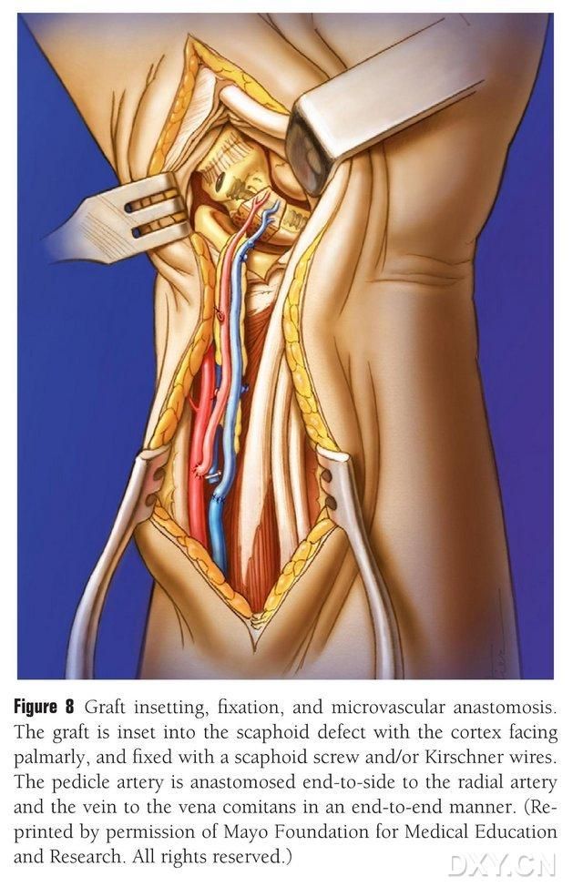
Pikitia 8. Te whakaurunga o te papa wheua, te whakau, me te anastomosis vascular. Ka āta whakauruhia te papa wheua ki te wāhi o te koha o te wheua navicular, ā, ka whakauhia mā te whakamahi i ngā tīwiri navicular tuwhera, i ngā pine Kirschner rānei. Kia tūpato kei te ōrite te taha metacarpal o te poraka wheua kua whakauruhia ki te taha metacarpal o te wheua navicular, kia pēhia rānei kia kore ai e pā. I mahia te anastomosis o te uaua papa vascular ki te uaua radial mai i te pito ki te pito, ā, i mahia te pito o te uaua ki te uaua hoa o te uaua radial mai i te pito ki te pito.
Te whakaora i muri i te pokanga
Āpirini waha 325 mg ia rā (mō te 1 marama), ka whakaaetia te amo i te taumaha o te peka e pāngia ana i muri i te pokanga, ka taea e te aukati turi te whakaiti i te mamae o te tūroro, i runga i te kaha o te tūroro ki te neke i te wā tika. Ka taea e te tautoko taha o te tokotoko kotahi te whakaiti i te mamae, engari kāore e hiahiatia te tautoko roa o ngā tokotoko. I tangohia ngā tuitui i ngā wiki e 2 i muri i te pokanga, ā, i puritia te Muenster, te maka roa o te ringa ki te koromatua mō ngā wiki e 3. Whai muri i tēnā, ka whakamahia te maka poto o te ringa ki te koromatua kia ora rā anō te whati. Ka tangohia ngā hihi-X i ia 3-6 wiki, ā, ka whakaūtia te whakaora whati mā te CT. Whai muri i tēnā, me tīmata mārire ngā mahi piko me te toronga kaha me te kore mahi, ā, me whakanui mārire te kaha me te auau o te whakakori tinana.
Ngā raruraru nui
Ko ngā raruraru matua o te hononga turi ko te mamae o te turi, te whara rānei o te io. I puta te mamae o te turi i roto i ngā wiki e ono i muri i te pokanga, ā, kāore i kitea he ngaronga o te rongo, he neuroma mamae rānei nā te whara o te io saphenous. Ko ngā raruraru matua o te ringaringa ko te kore hononga o te wheua uaua, te mamae, te pakari o te hononga, te ngoikore, te osteoarthritis haere whakamua o te ringaringa radial, o ngā wheua intercarpal rānei, me te mōrearea o te ossification heterotopic periosteal.
Te Tāruatanga Koreutu o te Wheua Kua Whakakīia ki te Vascular Condyle Femoral Medial mō ngā Nonunion Scaphoid me te Proximal Pole Avascular Necrosis me te Carpal Collapse
Wā tuku: Mei-28-2024


